
A radionuclide scan may be done for all sorts of reasons. A pet scan produces three dimensional colour images of your body using radionuclides.
 20 Pet Scan In Oncology
a pet scan uses what kind of radionuclides is important information accompanied by photo and HD pictures sourced from all websites in the world. Download this image for free in High-Definition resolution the choice "download button" below. If you do not find the exact resolution you are looking for, then go for a native or higher resolution.
Don't forget to bookmark a pet scan uses what kind of radionuclides using Ctrl + D (PC) or Command + D (macos). If you are using mobile phone, you could also use menu drawer from browser. Whether it's Windows, Mac, iOs or Android, you will be able to download the images using download button.
A bone scan is a common type.

A pet scan uses what kind of radionuclides. Pet scans take 20 to 30 minutes but you must wait about an hour while the tracer collects in the organ being studied. The system detects pairs of gamma rays emitted indirectly by a positron emitting radioligand most commonly fluorine 18 which is introduced into the body on a biologically active molecule called a radioactive. A radionuclide scan is an imaging technique that uses a small dose of a radioactive chemical isotope called a tracer that can detect cancer trauma infection or other disorders.
These chemicals localise to specific organs and tissues depending on the type of substance used and then emit small beams of radiation called gamma rays that can be detected by the gamma camera. A radionuclide is used which collects in areas where there is a lot of bone activity where bone cells are breaking down or repairing parts of the bone. In a cancer patient the cancer cells show up as denser areas on a pet scan due to the cells high metabolic rates in comparison with normal cells.
The dose of ionizing radiation from the tracer used in one pet scan for example typically exposes the patient to about 25 of the maximum allowable annual radiation exposure permitted for nuclear workers which is a very high limit to over 200 standardmodern medical chest xrays meaning a patient is getting exposed to the equivalent of. Positron emission tomography pet is a nuclear medicine functional imaging technique that is used to observe metabolic processes in the body as an aid to the diagnosis of disease. Pet stands for positron emission tomography.
So that only the scanner is required to perform a pet scan. Positron emission tomography pet. Pet scans show where cells are particularly active.
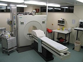
Devices used to scan patients who have been injected with small amounts of radionuclides and currently in use with other nuclear medicine procedures. Last updated on may 20 2019. Patient undergoing the process of getting a pet scan.
For a thyroid scan you take the radioactive tracer as a liquid or pill about 24 hours before the scan. See the separate leaflet called radionuclide scan isotope scan for more information on radionuclides. So a bone scan is used to detect areas of bone where there is cancer infection or damage.
The scan takes less than 30 minutes. The scan itself takes another hour or so. A radionuclide scan also known as a radioisotope scan is an imaging technique used to visualise parts of the body by injecting a small dose of a radioactive chemical into the body.
For example the pet scan shows exactly where the injected glucose is being used in the brain the heart muscle or a growing tumor.
 Spect And Pet
 By Nazli Gharraee April Ppt Video Online Download
 Characteristics Of Commonly Used Pet Radionuclides 2 8
 Radionuclide Imaging Dental Courses
 Radionuclide Imaging Dental Courses
 Positron Emission Tomography Wikipedia
 Spect And Pet
 Positron Emitting Radionuclides For Pet Download Table
 Commonly Used Single Photon Emitting Radionuclides
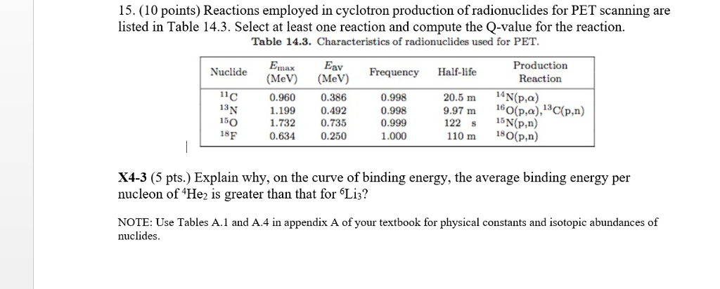 Solved 15 10 Points Reactions Employed In Cyclotron Pr
