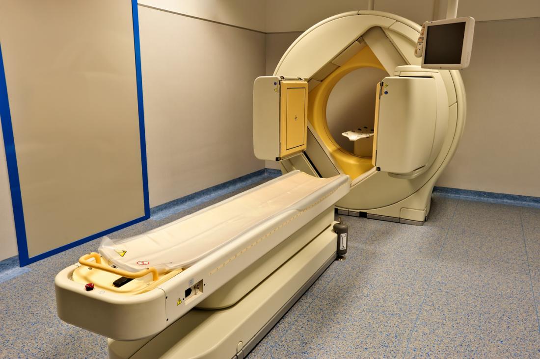
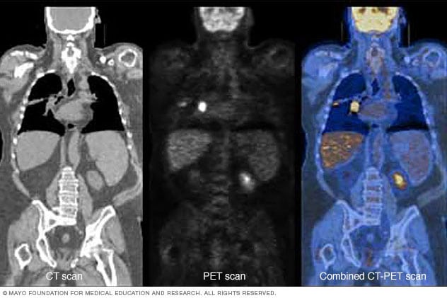
Machines that combine the pet and ct images called a petct are commonly used. The substances used in pet scanning are not associated with any side effects so you should feel no different after the scan.
what is used in a pet scan is important information accompanied by photo and HD pictures sourced from all websites in the world. Download this image for free in High-Definition resolution the choice "download button" below. If you do not find the exact resolution you are looking for, then go for a native or higher resolution.
Don't forget to bookmark what is used in a pet scan using Ctrl + D (PC) or Command + D (macos). If you are using mobile phone, you could also use menu drawer from browser. Whether it's Windows, Mac, iOs or Android, you will be able to download the images using download button.
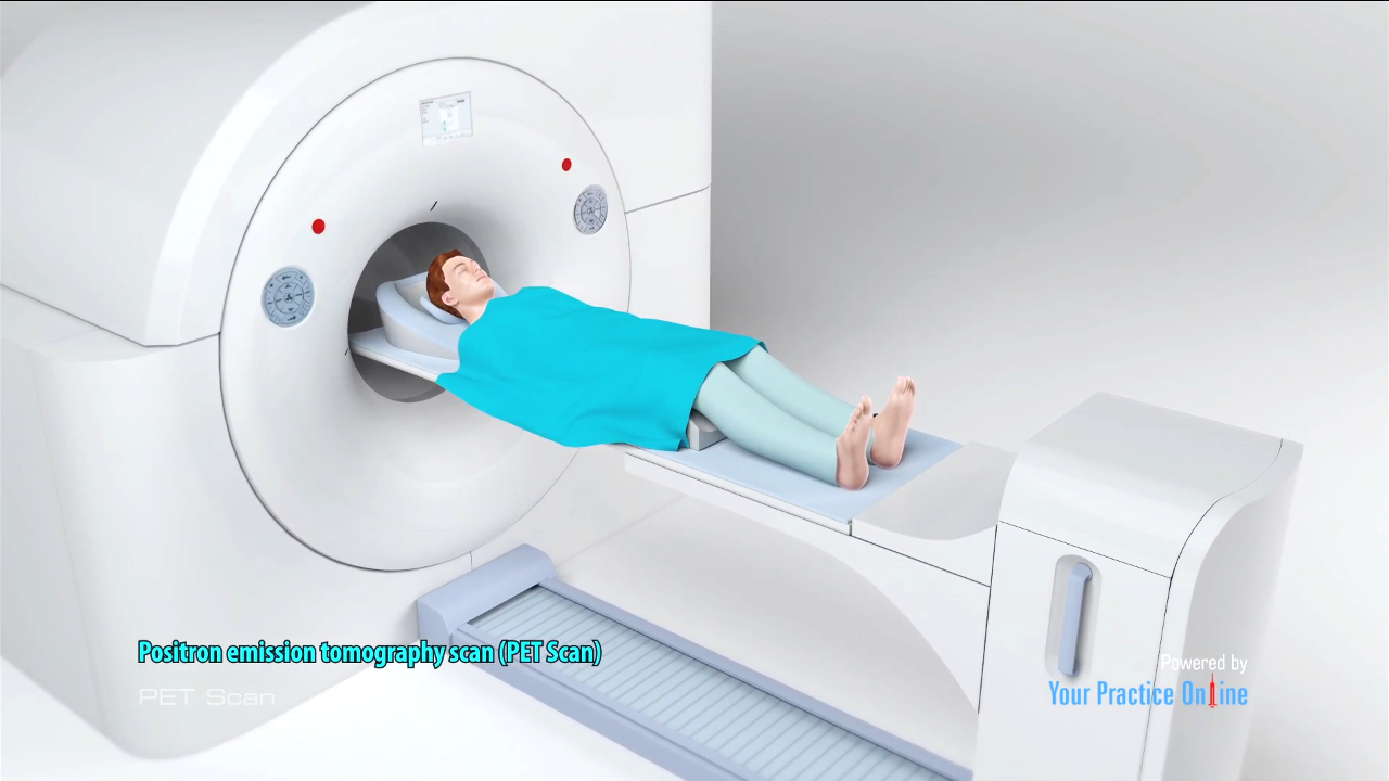
Positron emission tomography pet is a type of imaging technology used to evaluate how your tissues and organs work at the cellular level.

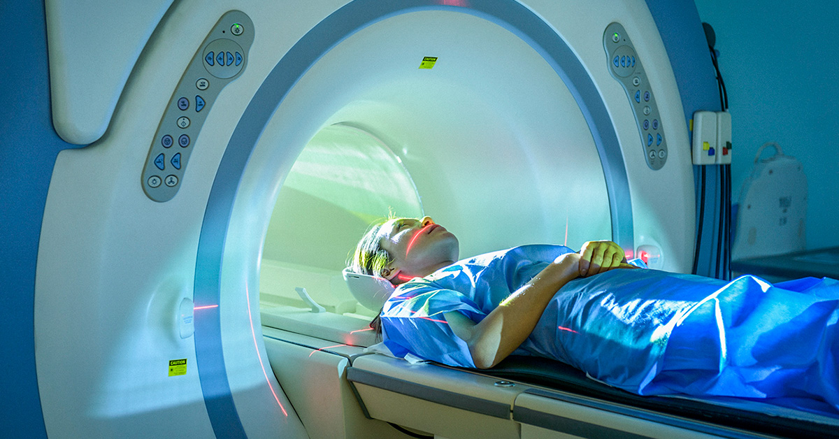
What is used in a pet scan. A pet scan is an imaging exam thats used to diagnose diseases or issues by how the body is functioning. Find out more about the procedure whether there are any risks and the difference between pet mri and ct scans. A positron emission tomography also known as a pet scan produces 3 d color images of the processes within the human body.
Millions of pet scans have been done around the world without complication. Well look into why its performed and how it compares to other tests. The pet scan uses a mildly radioactive drug to show up areas of your body where cells are more active than normal.
Pet stands for positron emission tomography. Unless your doctor tells you otherwise you can resume normal activities after a pet scan. This tracer is a glucose analog that is taken up by glucose using cells and phosphorylated by hexokinase whose mitochondrial form is greatly elevated in rapidly growing malignant tumors.
Its used to help diagnose some. They help your doctor measure blood flow oxygen use and more. Pet scans are often used to diagnose a condition or track how it is progressing.
A positron emission tomography pet scan is an imaging test that uses a special dye with radioactive tracers. Pet scanning with the tracer fluorine 18 18 f fluorodeoxyglucose fdg called fdg pet is widely used in clinical oncology. A pet scan is a very safe and routine procedure.
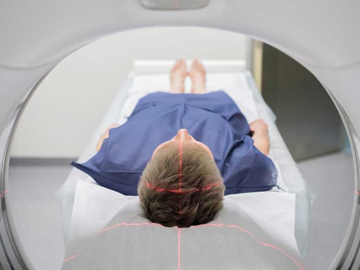
These tests show the structure of and blood flow to and from organs. The tracers are either swallowed inhaled or injected into your arm. It involves the injection of a short acting radioactive substance known as a radiotracer which is absorbed by biologically active cells.
It uses a special dye with radioactive tracers to help the machine capture changes in how the body is working such as how it absorbs sugar or how the brain is functioning. A positron emission tomography pet scan shows how organs and tissues are working. This is different than mri and ct scans.
Pet scans are a type of test that create 3 dimensional 3d pictures of the inside of your body.